Glaucoma

Understanding Glaucoma
Glaucoma is a category of eye disorders linked to dangerously raised internal eye pressure. The buildup in eye pressure can damage the eye’s optic nerve disrupting the transmission of visual information to the brain. Early stages of untreated glaucoma include a loss of your peripheral vision and progressed eye damage could eventually lead to blindness.
Most cases of glaucoma are not typically associated with pain and also produce no noticeable symptoms until vision loss occurs. Because of this, many cases go unnoticed until irreversible optical nerve damage has occurred along with a range of permanent vision loss.
There are two main types of glaucoma: open-angle glaucoma and closed-angle glaucoma. Open-angle glaucoma accounts for most cases. It is painless and the only signs are gradual loss of peripheral vision and optic nerve changes. Closed-angle glaucoma accounts for a small percentage of glaucoma cases. Closed-angle glaucoma symptoms include sudden eye pain, halos around lights, red eye, increased eye pressure, nausea and a sudden loss of vision. Closed-angle glaucoma is considered an eye emergency and should be handled by a professional to prevent permanent vision loss.
Routine eye exams include a screening for glaucoma using a machine called a tonometer. The tonometer is used to measure the pressure inside your eye. One of the most common tonometers use a puff sent onto the surface of the eye to test the internal pressure. Another method of glaucoma screening involved advanced imaging technology that monitors the eyes optic nerve over the course of your exams to ensure that no changes have occurred. Visual field testing is another way to determine loss of peripheral vision. It involves staring ahead while holding a button that will allow you to click when you see a flashing light in the edges of your vision. The test catches any blind spots that may develop as a result of damage to the optic nerve as a result of glaucoma.
Glaucoma treatment can involve surgery, or the use of lasers and/or medication depending on the progression. Eye drops with medication are one of the first forms of treatment used that aim to lower the internal pressure of the eye.
If you suspect you have glaucoma feel free to contact our office to schedule an eye exam with one of our doctors today. We will gladly answer any of your questions and provide you with the best treatment options available to you. If you experience any pain nausea or sudden vision loss, please call our offices immediately.
Optical Coherence Tomography (OCT)
OCT testing is a painless, non-contact procedure used to diagnose and monitor the progression of certain eye diseases like glaucoma. OCT imaging is one of the most helpful tools in diagnosing glaucoma, as it is capable of displaying images of fluids and other materials beneath the surface of the eye. This non-invasive test uses light waves that are dispersed just beneath the surface of the eye to produce high-resolution images of the biological tissues and fluids surrounding them. These cross-sectional images are used to measure the retinal nerve fiber layer. Thin layers can indicate the development or presence of glaucoma. Often, an eye doctor will take a baseline OCT imaging scan to use as a reference for future OCT scans. A progression in thinning of the retinal nerve layer can be a sign of glaucoma.
Visual Field Information
Visual field testing is used to obtain information about the visual field, including peripheral vision. During the test, patients stare at the center of a bowl-shaped instrument while lights flash or move in the peripheral field. Patients press a button each time they see a flash, allowing an ophthalmologist to identify patterns of vision loss that may be indicative of glaucoma. Excessive loss of peripheral vision indicates advanced optic nerve damage and requires immediate treatment to prevent further vision impairment or loss.
Specialized Testing
Glaucoma is a serious eye disease that can potentially cause total vision loss if not properly diagnosed, treated and managed. There are several different types of standard testing, such as visual field testing and dilated eye exams, that are used regularly to diagnose glaucoma. However, certain types of specialized testing may also be used to detect the presence of glaucoma, including optical coherence tomography (OCT) and pachymetry.
Pachymetry
A pachymeter is an ophthalmologic ultrasound instrument used to measure the thickness of the cornea. In less than a minute, sound waves are emitted to measure corneal thickness and to determine whether a patient’s cornea is normal, too thick or too thin. Normal thickness varies from person to person and that which is normal for one person may not be for another. Ocular pressure is correlated to corneal thickness, so pachymetry can be used to better develop a glaucoma treatment plan in patients with normal, thick or thin corneas.
We are currently enrolling in a Phase 3 FDA trial for glaucoma. Any interest, please call our study coordinator at 708-898-1858.
The surgeons at Multack Eye Care our proud to offer state of art customized Minimally Invasive Glaucoma Surgery (MIGS). With several options now available request an appointment today to discuss which option is best for your eyes.
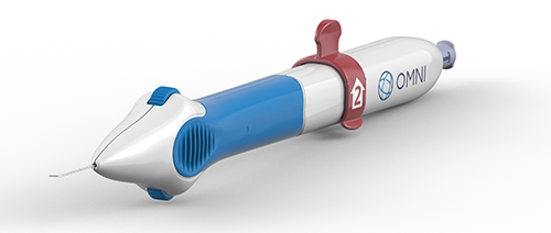
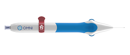
iStent Trabecular Micro-Bypass
Add an exciting new glaucoma technology to your cataract surgery.
iStent Trabecular Micro-Bypass
Technology has always played an important role in eye care. Today, almost every aspect of vision is connected to a product or procedure that wasn’t available even ten short years ago. The cataract surgery you are scheduled for is a good example of how innovations can make a difference. Every aspect of it utilizes recently developed technology that will help us improve your vision. Today, this includes managing your mild-to-moderate open-angle glaucoma: because now we are able to add another step to your cataract surgery that allows you to treat your open-angle glaucoma in a completely new way. This is important because once diagnosed, you and most patients like you will spend the rest of your lives putting one, two or even three different kinds of drops in every day. Unfortunately, all of these drops will not only be inconvenient, but potentially very expensive. The iStent Trabecular Micro-Bypass Stent is designed to reduce your eye pressure and you can have it done at the same time you have cataract surgery.
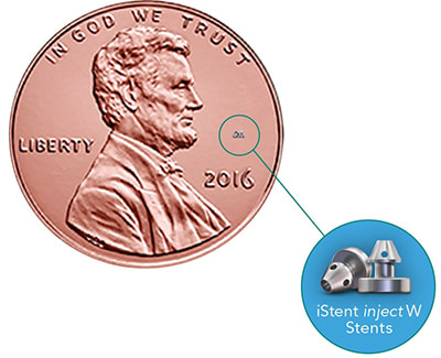
iStent®: the world’s smallest medical implant delivers big results in mild-to-moderate open-angle glaucoma.
While mild-to-moderate open-angle glaucoma is very common, many people are unaware of their condition, especially in the early stages, when their vision may be unaffected. In many people, open-angle glaucoma is characterized by an increase in the intraocular pressure (IOP) of your eye. This pressure is caused by the buildup of fluid within the eye. Too much fluid raises pressure, which can cause the gradual loss of vision. And while glaucoma moves slowly, its damage is irreparable.
The world’s tiniest medical device—iStent Trabecular Micro-Bypass—is 20,000 times smaller than the intraocular lenses (IOL) used in your cataract surgery. But the size of iStent Trabecular Micro-Bypass is only part of its story. By increasing the eye’s ability to drain fluid, this technology is designed to reduce the pressure in your eye.
In a U.S. clinical study, 68% of glaucoma patients who received iStent Trabecular Micro-Bypass remained medication free at 12 months while sustaining a target IOP of ≤ 21 mm Hg vs. only 50% of patients who underwent cataract surgery alone.
iStent works like the stents used to prevent heart attacks and strokes. When blood vessels get clogged, a stent creates access to the vessel flow. While a highly innovative technology, how iStent works is elegantly simple:If you have glaucoma, over time the eye’s natural drainage system becomes cloggediStent creates a permanent opening through the blockage to improve the eye’s natural outflowRestoring this mechanism lowers and controls pressure within the eye
