Technology
Utilizing State-of-the-Art-Technology For Vision Care
At Multack Eye Care we mean it when we say “Come See the Difference Today” and “Most Advanced Technology in the Nation”. This ideal drives everything we do and every decision we make – investing in the latest technology innovations, being a part of cutting-edge vision research studies, and training and hiring the most experienced and proven surgeons and staff in all of eyecare. At the center of our vision is the customized patient experience – our excellence in technology, research, and surgical results is the foundation of our care, and our commitment to patient education, listening, and fully customized treatment plans is the way we strive to make our focus, your best vision! Here are just some of our State of the Art Technology:
Digital Imaging
We believe in accuracy and superior outcomes and with that means investing in technology that helps us achieve that. The most modern cataract surgical equipment provides our surgeons with real time image measurements and digital overlays for the accurate guidance in cataract surgery available.
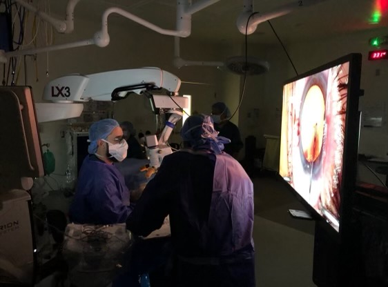
Surgical Microscope
The LuxOR® Ophthalmic Microscope advances visualization for our surgeons with personalized illumination, giving you and the surgeon confidence throughout the procedure.
Ngenuity 3D Visualization System
Allows our surgery 5X more depth of field, and crisper focus across an expanded space to allow them during surgery to have a deeper range for detail.
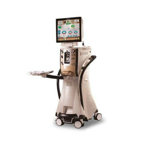
Active Sentry Centurion
Ultrasonic phacoemulsification has long been the “gold standard” for cataract surgery. In phacoemulsification surgery, a small ultrasonic probe is inserted into a very small incision on the edge of the eye. This probe gently breaks the cloudy lens into tiny pieces and suctions the cataract out of the eye. The Centurion offers a new method that uses both ultrasound and mechanical oscillation to help break up the cataract faster, and allows the surgeon greater control of lens tissue than traditional ultrasound.
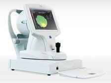
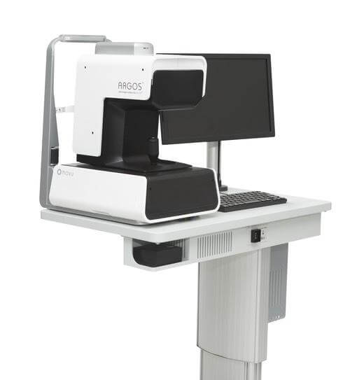
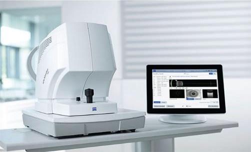
Cataract measurement/imaging technology: Biometers, Topographer, and Tomographer
Multack Eye Care is one of the only Ophthalmology Clinics to own all the latest measuring devices available. We invested in modern technologies to achieve multiple and accurate images, scans, maps, and measurements of the eye, from front to back and everything in between, before cataract surgery.

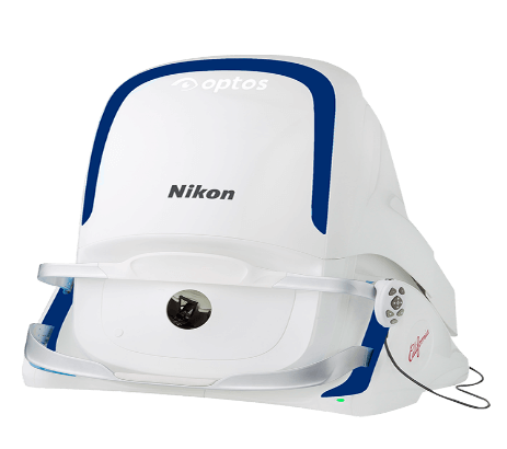
Retinal (Fundus) Cameras: Ultra-Wide Field
Diagnosing Retinal disease as soon as possible is a priority at Multack Eye Care. Therefore we reinvest into the latest and most advanced technology to best help us achieve earlier diagnosis. Here is why we purchased these specific technologies.
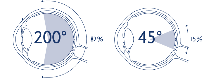
The peripheral retina is the site of pathology in many ocular diseases, including diabetic retinopathy (DR), retinal vein occlusion (RVO), uveitis, retinal vasculitis, and rhegmatogenous retinal detachment (RRD). Careful clinical examination of the peripheral retina with scleral indentation is important in clinical decision-making when there is a need for additional testing and objective, reliable documentation of findings.
Diagnostic imaging techniques play an increasingly prevalent role in assessment of the retinal periphery. Ultra-widefield (UWF) imaging has changed the way ophthalmologists evaluate a patient’s fundus. Older technology only captures up to 30 to 45 degrees of the retina. Both our Ultra-Wide Field technologies capture over 200° up to 267 ° making these the absolute best fundus cameras available.
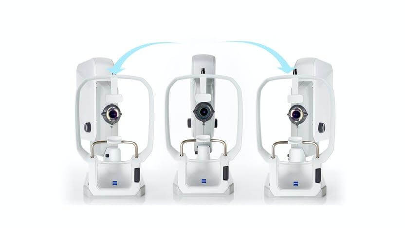
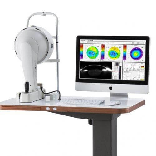
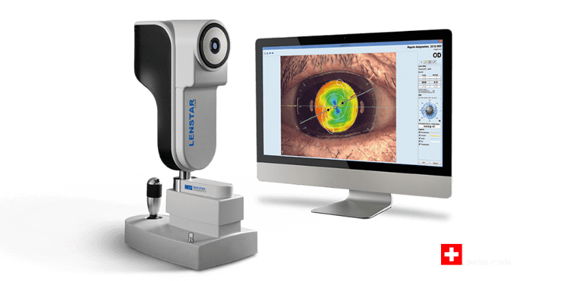
Topographer and Tomographer
The cornea is the outer layer of the eye, responsible for about 70 percent of the eye’s focusing power. A corneal topography test provides detailed 3D maps of the cornea’s shape and curvature and enables detection of corneal diseases, and irregular corneal conditions, such as swelling, scarring, abrasions, deformities, and irregular astigmatisms.
Corneal tomography is a more advanced diagnostic tool because its Scheimpflug imaging takes photographs of the anterior and posterior cornea. As a result, the posterior cornea can be visualized, which can be abnormal in early onset keratoconus or early post-LASIK ectasia.